CEXCA – Biopsy
BIOPSIES
FINE NEEDLE ASPIRATION BIOPSY
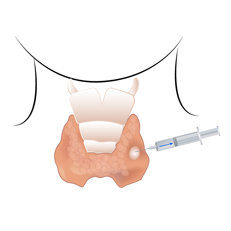
Fine needle aspiration (FNA) biopsy is the first diagnostic method for the study of thyroid nodules or some neck masses. The thyroid is a gland found in the anterior part of the neck, just below the Adam’s apple, and its function is to produce thyroid hormones which regulate the body’s metabolism.
A small needle is inserted into the nodule to obtain enough material to help determine if the nodule is benign or malignant.
The procedure is carried out in an ambulatory and has a diagnostic accuracy of over 95%. The needle is so thin that the procedure dispenses with the use of local anesthetics. If the nodule is palpable or measures more than two centimeters, the procedure is performed directly by the surgeon. If the nodule is not palpable or less than 2 cm, the biopsy should be performed under ultrasound guidance, usually by a radiologist.
The person lies on their back with their neck extended. The puncture site is sterilized with alcohol and the needle inserted, where two or three entry and exit movements are made, and the needle finally removed. Gentle pressure is applied to the site for two minutes. The procedure takes approximately one minute. Occasionally, a second puncture is needed if the first did not produce enough material.
No preparation is necessary. Most people report mild pain which disappears after a short while. There is an extremely minimal risk of bruising, which is usually treated with common analgesics and local measures.
The sample obtained is processed by an expert pathologist, who takes approximately two weeks. The result must be interpreted by a head and neck surgeon or an endocrinologist.
ORAL CAVITY BIOPSY
An open biopsy is the first diagnostic method for the study of masses found in the mouth.
A small sample is collected from the mass in sufficient amount to help determine if the nodule is benign or malignant.
The procedure is carried out in an ambulatory with diagnostic accuracy of over 95%. Topical anesthesia is usually applied in small amounts.

The person stays seated. The biopsy site is sterilized with an iodine solution, and topical anesthesia is then applied and allowed to act for a few minutes. A sample is then collected with the use of a special biopsy forceps. The patient will feel pressure and a jerking sensation. Gentle pressure is then applied to the site for two minutes to control bleeding. This procedure takes approximately one minute. Usually, a second and even a third biopsy may be needed to obtain enough material. After the biopsy, the patient presents a painful and sensitive peel-off for a few days.
No preparation is necessary. Most people report mild, short-term pain, and the minimal bleeding is controlled with the application of pressure. There is an extremely minimal risk of bruising or infections, usually treated with common analgesics and local measures. The sample obtained is processed by a pathologist and takes about two weeks to process. A head and neck surgeon should interpret the result.
